Adding product to the cart!
Added to the cart!
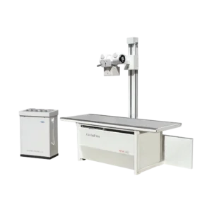
ARI-30MX X-Ray
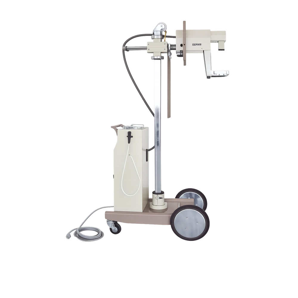
This portable X‑ray unit provides clear imaging for early detection of mammary pathological changes, distinguishing benign from malignant tumors with high accuracy. Its mobile design allows bedside diagnostics and nondestructive inspection of both metal and nonmetal materials.
Description
- It is used to diagnose early mamma pathological changes
- Using the Mo. target X-ray tube can show the pathological changes details.
- A nipple areola cuticles fat galactophore canals glandular tissue connective tissue and blood vessels
can be seen in the picture.
- It has high correctness for distinguishing benign tumor and malignant tumor.
- It can be moved to sickroom to photograph beside bed.
- The unit can also be used to find foreign matters in human body to do nondestructive inspection of right
metal and nonmetal materials.
- The movement of camera head mounting: vertical 630 mm
- Adapt to different part of human Rotation ±180°
- The camera head mounting horizontal range.160 mm
- Rotating angle: module of the camera, head mounting and the pole:180°
- Specification of X-ray tube/focal spot value:XD7 – 1.05/35 1mm×1 mm
- Stationary anode: single focus
Technical Parameters
| Parameter |
Value |
| Tube Current |
36 kV |
| Tube Voltage |
1.5 mA |
| X-ray Bulb Focus |
0.3 mm |
| X-ray Bulb |
D-081 |
| Power |
250 AV |
| Gross Weight |
55 KG |
| Current |
30 mA |
| Voltage |
34 kVp |
| Time |
2 s |
| Power Supply Voltage |
180–240 V |
| Frequency |
50 Hz |
| Power Not Lower Than |
1.3 kVA |
| Range of the Timer |
0.4 s |
| Film Size |
127 mm × 178 mm |